

What is patella instability?
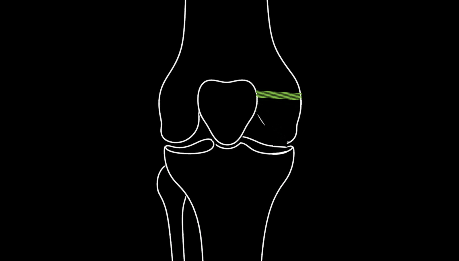
The kneecap, or patella, sits in a groove at the front of the thigh bone and slides up and down that groove as the knee bends and straightens. Several structures keep it there: the shape of the groove itself, the alignment of the leg, the muscles around the knee, and a ligament on the inner side called the medial patellofemoral ligament, or MPFL.
When the kneecap comes fully out of the groove, that is a dislocation. When it slips partly out and returns, that is a subluxation. Both are described as instability.
The first dislocation is usually caused by a twisting movement or a direct blow, often during sport. It is typically painful and obvious, with immediate swelling. The kneecap sometimes relocates itself as the knee straightens.
The problem is what happens afterwards. The MPFL is damaged in the great majority of first-time dislocations, and it heals poorly. Once it has been stretched or torn, the kneecap is more likely to come out again.
Why does it keep happening?
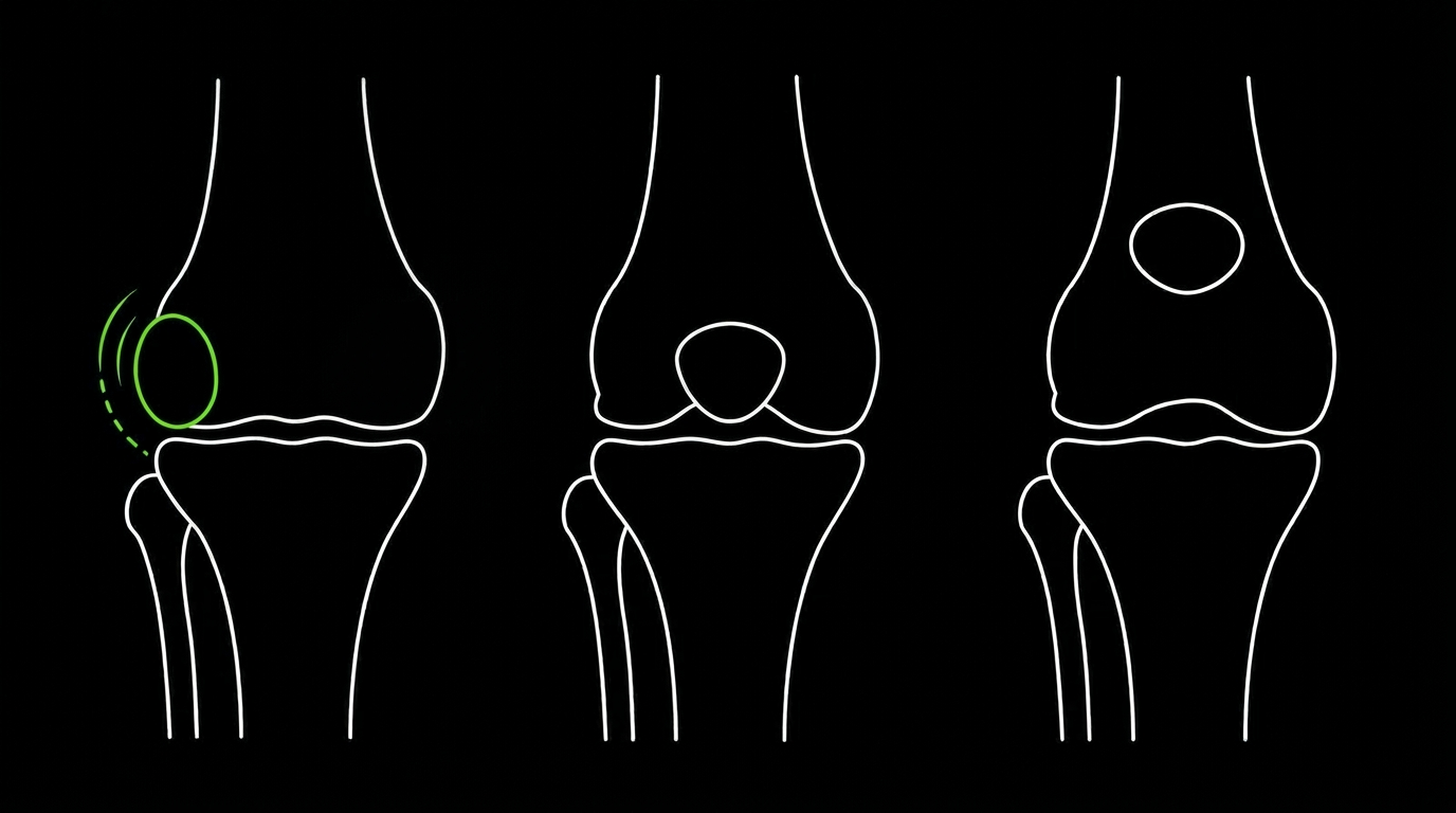
Some people dislocate once after a significant injury and never have another problem. Others dislocate repeatedly, sometimes with very little force, and this is usually because of the underlying shape and alignment of the knee rather than the original accident.
Factors that make recurrence more likely include:
Trochlear dysplasia. The groove that the kneecap runs in is shallower than normal, so there is less to hold the kneecap in place.
Patella alta. The kneecap sits higher than normal, so it engages the groove later in the range of movement and is less stable in the early part of bending.
Increased TT-TG distance. The pull of the quadriceps and the position of the tendon attachment on the shin bone are offset, drawing the kneecap outwards.
Generalised ligament laxity. Naturally flexible joints throughout the body.
Rotational alignment. The way the thigh bone and shin bone are rotated relative to each other.
Muscle weakness or poor control, particularly of the quadriceps and hip muscles.
These factors matter because they determine what treatment is appropriate. Reconstructing the ligament alone may not be enough if the underlying alignment is significantly abnormal. Assessment therefore involves X-rays and usually an MRI or CT scan to measure these features before any decision about surgery is made.
Who is patella instability surgery suitable for?
Surgery is usually considered when:
- The kneecap has dislocated more than once
- Instability continues despite a proper course of physiotherapy
- The knee gives way or the kneecap slips during everyday activity or sport
- A fragment of bone or cartilage was broken off during a dislocation
- Fear of dislocation is restricting what you can do
- Imaging shows anatomical factors that make further dislocations likely
After a first dislocation
A first dislocation is usually treated without surgery. Physiotherapy to restore strength and control, with a period of bracing or support, is the normal starting point. Surgery is generally reserved for those who dislocate again, or for those with a loose fragment inside the knee that needs dealing with.
There are exceptions. Where imaging shows significant anatomical risk factors, or where a fragment of bone or cartilage has been broken off, earlier surgery may be recommended.
Who it is not suitable for
If your kneecap has dislocated once and your knee now feels stable, surgery is unlikely to be advised. If you have anterior knee pain without instability, patella stabilisation surgery is not the answer and may not help the pain. If you have not yet completed a proper rehabilitation programme, that usually comes first.
Mr John Lynch will assess your knee, review your imaging and discuss whether surgery is likely to help before recommending it.
What surgery is involved?
There is no single operation for patella instability. The procedure is chosen to address the specific reason your kneecap is unstable, which is why the imaging assessment matters.
MPFL reconstruction
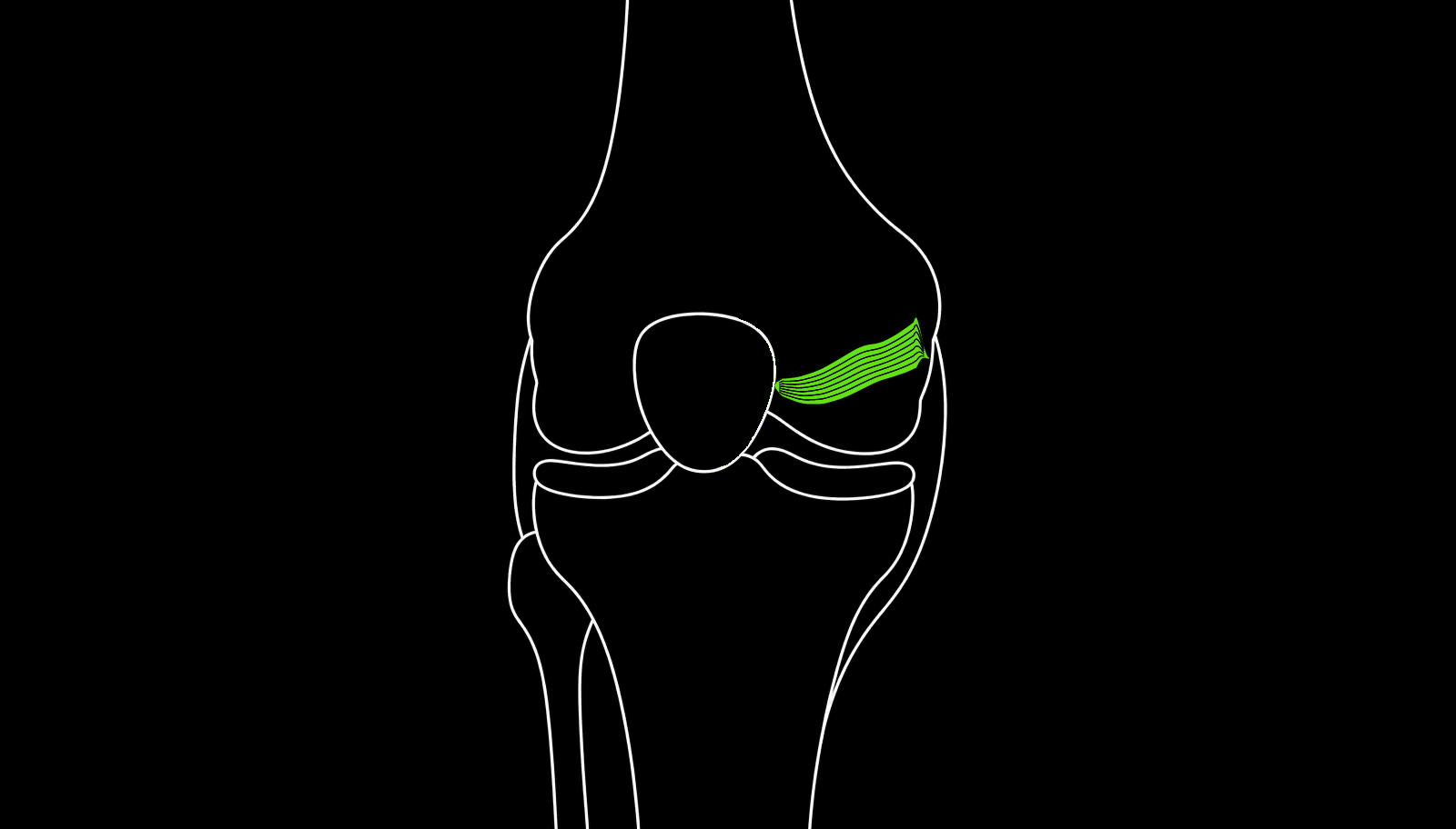
The most common operation. The damaged ligament on the inner side of the kneecap is replaced with a graft. The graft is attached to the kneecap at one end and to the thigh bone at the other, restoring the restraint that stops the kneecap sliding outwards.
It is carried out through small incisions, often combined with an arthroscopy to inspect the joint and deal with any cartilage damage. Most patients go home the same day or the following day.
Tibial tubercle osteotomy
Where the alignment is significantly abnormal, the bony attachment of the patellar tendon on the shin bone is moved and refixed with screws in a better position. This changes the direction of pull on the kneecap. It is a larger operation, with a longer recovery, and it is often combined with MPFL reconstruction.
Trochleoplasty
Where the groove itself is severely misshapen, it can be deepened surgically. This is a specialised procedure reserved for a small number of cases with severe trochlear dysplasia.
Lateral release or lateral lengthening
Loosening tight tissue on the outer side of the kneecap. This is rarely done on its own and is generally used alongside other procedures where tightness is contributing to the problem.
Removing or fixing loose fragments
Where a dislocation has broken off a piece of bone or cartilage, this may need fixing back or removing, sometimes urgently.
Mr John Lynch will explain which procedure, or combination, is appropriate in your case and why.
What is recovery like?
Recovery depends considerably on which operation you have. The timescales below relate to MPFL reconstruction. If a tibial tubercle osteotomy is carried out as well, recovery is longer and weight-bearing is usually restricted while the bone heals.
The first two weeks
You will be on crutches, often with a brace controlling how far the knee bends. The priorities are settling swelling, regaining full straightening, and getting the quadriceps working again. Pain is generally more manageable than after larger knee operations.
Weeks two to six
Bending is progressively increased, usually according to a set schedule. Crutches are discarded as walking normalises. Physiotherapy focuses on restoring movement and on the muscles that control the kneecap.
Six weeks to three months
Strength work becomes the priority, including the hip and core muscles as well as the quadriceps. Static cycling, controlled gym work, and progressive loading. Most people return to desk work well before this point.
Three to six months
Running is reintroduced when strength and control allow. Sport-specific work follows, progressing from straight-line movement to changes of direction.
Six to nine months
Return to pivoting and contact sport is usually possible during this period, guided by strength testing and confidence rather than by a fixed date.
What to expect from the result
The aim of surgery is a kneecap that stays where it should. Most people regain a stable knee and return to their previous activities.
Confidence often takes longer to return than physical strength. Many people are cautious about the movement that caused the original dislocation for some time after they are physically ready.
Some people continue to have aching at the front of the knee, particularly on stairs or after prolonged sitting. Stabilisation surgery addresses instability reliably; it is less predictable for pain.
What are the risks?
- Recurrent dislocation or continued instability despite surgery
- Ongoing pain at the front of the knee
- Stiffness or loss of full bending
- Fracture of the kneecap, a recognised risk where tunnels are made in the patella
- Infection
- Blood clots in the leg or lungs
- Numbness around the incisions, often permanent
- Discomfort from screws or fixation devices, occasionally requiring removal
- Weakness of the quadriceps
- Where a tibial tubercle osteotomy is performed, additional risks including fracture and delayed bone healing
- Risks associated with anaesthesia
Mr John Lynch will discuss your individual risks with you, based on your anatomy, your history and the procedure planned.
How effective is patella stabilisation surgery?
The evidence supports surgery where dislocations are recurrent.
Following a first dislocation treated without surgery, published recurrence rates range from around 15% to 44%.
One systematic review comparing treatments after a first-time dislocation found redislocation in around 7% of those who had early MPFL reconstruction, compared with around 30% of those treated with rehabilitation alone.
A review of MPFL reconstruction with a minimum five-year follow-up reported an overall redislocation rate of 5.75%, with significant improvements in patient-reported knee scores.
Numbers from published studies describe groups of patients, not individuals. Your own likely outcome depends on your anatomy, which procedure is appropriate, and how you rehabilitate.
What are the alternatives?
Physiotherapy. A structured programme targeting the quadriceps, the hip muscles and control of the knee during movement. This is the first treatment after a first dislocation and helps many people avoid surgery altogether.
Bracing and taping. A brace or taping can improve confidence and give some support, particularly during sport. It does not correct the underlying cause.
Activity modification. Avoiding the movements that provoke instability. This is a reasonable choice for some people, though it can mean giving up sports.
Treating the pain rather than the instability. Where the main problem is anterior knee pain rather than the kneecap coming out, the treatment is different and usually does not involve stabilisation surgery.
Watchful waiting. Some people dislocate once and never again. Where instability is not recurring, monitoring is often more sensible than operating.
What does patella instability surgery cost?
Patella stabilisation surgery is available on a self-pay basis or through private medical insurance. Costs vary depending on which procedure is required. Please contact us for current fees, to discuss what is included, and to check which insurers are recognised. Physiotherapy forms a significant part of the treatment, so it is worth establishing at the outset how much is covered.
Book a consultation
To arrange an appointment, please call us or use the enquiry form.